
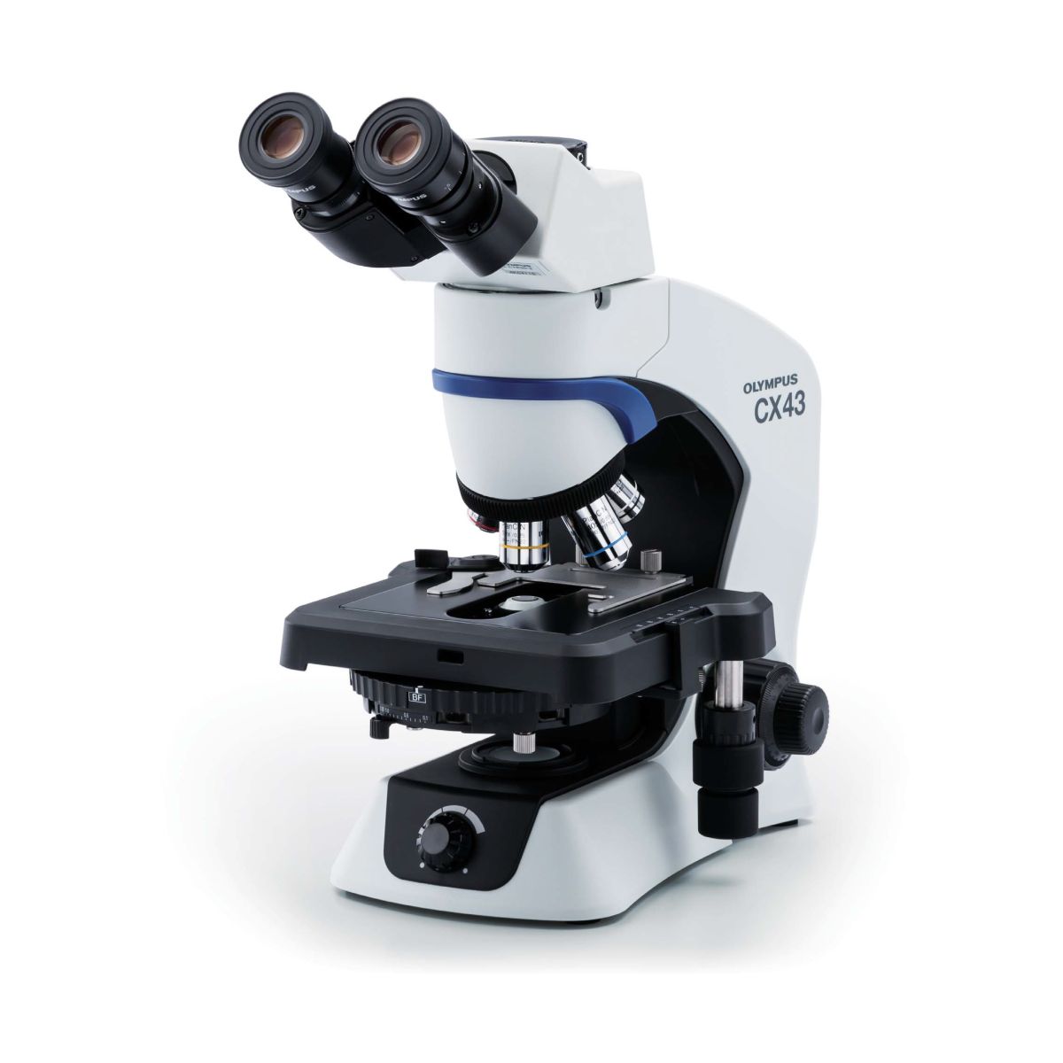
Olympus cx43 microscope: characteristics and features of the device
The best device in its line. Designed for the purpose of complex work. Due to its ergonomics, it is well suited for work in small laboratories, schools, mobile laboratories where it is possible to carry out simple research.
Features of the device
The main features of the microscope include:
- entered in the register of the Russian Federation of medical devices;
- has an anti-fungal coating;
- there is a led indicator illuminator using 2.5 W of electricity;
- has a universal insert for dark-field microscopy;
- there is a diopter adjustment for the left eyepiece tube;
- equipped with front lenses with 100x and 40x lenses. To move the lens, you just need to lightly touch it. When you move the lens to a position, it snaps into position. Any lenses from the Olympus UIS2 series are mounted on the device;
- electric illuminator CX43-RFAB is available, you can manually adjust the brightness in it;
- There is a drawing attachment;
- support for UIS2 optics - polarizing lenses;
- field of vision - 2 cm, also, the indicator of the field of view - FN22, is considered good for clinical diagnosis;
- if necessary, you can immediately observe, photograph and record video at the same time;
- for U-GAN analyzers there is an intermediate insert called CX3-KPA. They check urine for foreign bodies, etc.
- for a stage with a holder, one glass slide is placed. Also, it is equipped with 2 glasses. To change the slides without harming the microscope, a rubber backing can be fitted. There is a clamping holder with which you can move the sample with your fingers;
- service life of about 60,000 hours. If translated into more understandable numbers, then this is about 17 years;
- protected from static electricity;
- can be zoomed in smoothly and quickly with the rotating tip located at the bottom;
- turret condenser with six positions. There are three sliders for phase contrast;
- storage at: from - 26 ° С to 66 ° С of the environment, humidity up to 91%.
Main characteristics
1. Lighting system - located in a tripod. Led indicator light source for 2.6 watts.
2. Objective revolver - the revolver itself is 5 positions with an internal bias. That is, it fixes 5 lenses.
3. Focusing - coarse and fine focusing of coaxial screws. When the coarse focusing screw rotates, its area of movement becomes - 3.8 cm. On coarse focusing, the effort is adjusted.
4.Fluorescent light source - LED illuminator. 469 nm. Excellent image clarity, without any distortion.
5. Subject table - size 210 x 155 mm. Abscis and ordinate travel - 75 x 53 mm. The movement of the drug carrier is blocked if necessary. Reliable drug holder that can still be opened easily. Rubberized grips for control.
6. Tube - tilt 30 °. Coating - Anti-fungal. The distance between the pupils is 49-74 mm.
7. The Abbe condenser is a seven-position universal condenser. There is a retainer in brightfield mode. Iris lock is used to lock the contrast. It also has inserts for light and dark fields, and phase contrast. For phase contrast, special PH lenses are needed.
8. Contrast - dark and light field, fluorescence, polarization.
9. Optical system - UIS2.
10. Connection to the network - from 99.9-240V, frequency in the range - 50/60 Hz, voltage 0.3-0.5A.
The microscope is easy to use and relatively inexpensive. This device has no drawbacks, so it will serve its owner for many years. Suitable for use by novice scientists in scientific laboratories.